top of page
Interventional.jpg

Jackpersad & Partners Inc.

Full Services 

We pride ourselves in offering professional and exceptional care and service to both our referring doctors and patients.

Mammography Unit 2.jpg

MAMMOGRAPHY

Mammography is a specific type of imaging that uses a low-dose x-ray system to examine breasts and is used to aid in the early detection and diagnosis of breast diseases in women.

BREAST BIOPSY

A Breast Biopsy is where a special needle is inserted into the breast to take a small sample of breast tissue from an area of concern so that it can be sent to a laboratory for testing. An abnormality may have been identified on a scan or X-ray image or may have been felt as a lump.

Breast Biopsy edited.jpg
IMG_8946.JPEG

ULTRASOUND

Ultrasound includes sound waves in the frequency range of Twenty Thousand Hertz or cycles per second, which cannot be heard by the human ear. Ultrasound in medical imaging uses frequencies in the 2-12 million hertz range.

BONE DENSITOMETRY (BMD)

Bone densitometry, also called dual-energy x-ray absorptiometry (DEXA) uses low dose xrays to produce images of the bones. It is most commonly performed on the lumbar spine and hips but sometimes also includes the distal forearm. There is good correlation between the amount of bone measured, and bone strength.

Screenshot 2022-03-16 145145.jpg
Image by Umanoide

GENERAL X-RAYS

An x-ray (radiograph) is a non-invasive medical test that helps physicians diagnose and treat medical conditions. Imaging with x-rays involves exposing a part of the body to a small dose of ionizing radiation to produce images of the body.

COMPUTERISED TOMOGRAPHY (CT)

A CT scan is a diagnostic imaging procedure that combines x-ray technology and computer-generated cross-sectional images or "slices" of the body. A CT scan shows detailed images of any part of the body, including bones, muscles, fat, blood vessels and organs.

IMG_8923.JPEG
Interventional.jpg

INTERVENTIONAL RADIOLOGY

Interventional Radiology (abbreviated IR or sometimes VIR for vascular and interventional radiology) is a subspecialty of radiology in which minimally invasive procedures are performed using image guidance. 

MAGNETIC  RESONANCE IMAGING

We have highly trained and experienced M.R. technicians and radiologists.
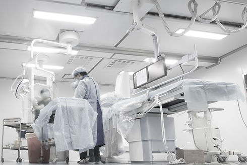
With this sophisticated equipment at our disposal, we can ensure that patients are imaged as swiftly as possible, and an expert MR radiologist is always at hand to provide the patient with reassurance and explanation of how the scan is progressing.

P1070522-min.jpg
Nuclear Medicine 3.jpg

NUCLEAR MEDICINE

  • Nuclear Medicine is a branch of medical imaging that uses the nuclear properties of matter in diagnosis and therapy.

  • Nuclear Medicine uses radionuclides/radioisotopes or radiopharmaceuticals that are either injected into the vein or intradermally, ingested with food or liquids or inhaled in gaseous form.

FLUOROSCOPY STUDIES

Fluoroscopy is a type of medical imaging that depicts a continuous x-ray image on a monitor. Doctors use fluoroscopy to visualise structures inside the body in real-time during medical procedures. 

Shifa Fluoroscopy.jpg
PET CT.jpg

PET CT

PET is an abbreviation for Positron Emission Tomography and is performed in the Nuclear Medicine department. It involves the use of a radioactive substance called F18-FDG (Fluorodeoxyglucose) which traces metabolic activity. 

SPECT CT

SPECT is a scintigraphic technique in which a computer-generated image of local radioactive tracer distribution in tissues is produced through the detection of single-photon emissions from radionuclides introduced into the body that is able to provide true three-dimensional (3D) information. T

Nuclear Medicine SPECT CT.jpg
Mammo - Hillcrest.jpg

TOMOSYNTHESIS

Tomosynthesis is an imaging technique in which multiple X-rays of the breast are taken from a discrete number of angles. These cross-sectional images are used to reconstruct 3-D images of the breast being imaged. 

bottom of page