
Acerca de


An x-ray (radiograph) is a non-invasive medical test that helps physicians diagnose and treat medical conditions. Imaging with x-rays involves exposing a part of the body to a small dose of ionizing radiation to produce images of the body. X-rays are the oldest and most frequently used form of medical imaging.
X-rays are a form of radiation like light or radio waves. X-rays pass through most objects, including the body. Once it is carefully aimed at the part of the body being examined, an x-ray machine produces a small burst of radiation that passes through the body, recording an image on photographic film or a special detector.
The equipment typically used for bone x-rays consists of an x-ray tube suspended over a table on which the patient lies. A drawer under the table holds the x-ray film or image recording plate. Sometimes the x-ray is taken with the patient standing upright, as is the case with chest x-rays.
A portable x-ray machine is a compact apparatus that can be taken to the patient in a hospital bed or the emergency room. The x-ray tube is connected to a flexible arm that is extended over the patient while the x-ray film holder or image recording plate is placed beneath the patient.
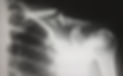
Different parts of the body absorb x-rays differently. Dense bone absorbs more radiation while soft tissue, such as muscle, fat and organs, allow more of the x-rays to pass through them. As a result, bones appear white on the x-ray, soft tissue shows up in shades of grey and air appears black.

Until recently, x-ray images were maintained as hard film copy (much like a photographic negative). Today, most images are digital files that are stored electronically. These stored images are easily accessible and are frequently compared to current x-ray images for diagnosis and disease management.
The radiographer, an individual specially trained to perform radiology examinations, positions the patient on the x-ray table and places the x-ray film holder or digital recording plate under the table in the area of the body being imaged. When necessary, sandbags, pillows or other positioning devices will be used to help to maintain the proper position. A lead apron may be placed over your pelvic area to protect you from radiation.
You must remain very still while the x-ray is being taken. You may be asked to hold your breath for a few seconds, to reduce the possibility of a blurred image.
You may be repositioned for another view and the process is repeated. Two or three images (from different angles) will typically be taken.
An x-ray may also be taken of the unaffected limb, or of a child's growth plate (where new bone is forming), for comparison purposes.
When the examination is complete, you may be asked to wait until the radiologist determines that all the necessary images have been obtained.
A bone x-ray examination is usually completed within 15 to 30 minutes.